Dr. Domb is a nationally recognized orthopaedic surgeon specializing in sports medicine, regenerative medicine, and arthroscopic surgery of the shoulder. Dr. Domb is an expert in arthroscopic surgery of the shoulder, adept in specialized techniques including arthroscopic rotator cuff repair.
- Shoulder Basics
- Shoulder Injuries & Conditions
- Non-surgical & Regenerative Medicine
- Surgeries Performed by Dr. Domb
Shoulder Anatomy
- The shoulder is a sophisticated, “ball and socket” joint comprised of three bones (clavicle, humerus and scapula) and four individual joints (glenohumeral, acromioclavicular, sternoclavicular, and scapulothoracic). Together, these are known as the shoulder girdle.
- The clavicle, also known as the collar bone, is the only bony attachment between the upper limb and the trunk. It articulates on the medial end with the sternum and on the lateral end, with the acromion of the scapula. Furthermore, the clavicle forms the anterior portion of the shoulder girdle.
- The glenoid cavity is the shoulder structure that serves as the “socket.” The humeral head fits into the shallow glenoid cavity, providing the “ball” to make up the ball and socket joint. Compared to the hip “socket,” or acetabulum, the lack of depth in the cavity affords the humerus complete rotation about the joint. However, due to its skeletal structure, the shoulder is relatively unstable. The rotator cuff muscles, the labrum, and glenohumeral ligaments provide joint stability.
- The scapula is a large, flat, triangular shaped bone with three distinct processes: the acromion, spine and corocoid process. The articulation formed between the acromion and the clavicle makes up the acromioclavicular (AC) joint. The scapula articulates laterally with the humeral head at the glenoid cavity and glides along the back of the chest during arm extension. The coracoid process is on the anterior side and serves as an attachment for ligaments and muscles.
- The shoulder also contains a significant amount of soft tissue. It is comprised of several muscles, tendons, ligaments and cartilage. The rotator cuff is a group of four muscles and tendons that originate on the scapula and attach distally on the tuberosities of the humerus. These muscles provide stability by keeping the humeral head centrally located in the glenoid cavity, or fossa, by surrounding the joint, creating a cuff to support movement of the ball and socket. The rotator cuff is also essential in moving the arm as it allows the arm to be raised overhead, in front of the body or to the side.
- In addition to muscles and tendons, ligaments also provide stability to the shoulder by forming the joint capsule that surrounds the glenohumeral joint. The labrum is a ring of elastic cartilage found within the shoulder socket, surrounding the edge of the glenoid fossa. It acts as a container for the humeral head, deepening the socket to maintain stability of the shoulder. It creates a suction seal to further act as a stabilizer of the shoulder joint.

Rotator Cuff Tears
The rotator cuff is a group of four muscles and tendons that originate on the scapula and attach distally on the tuberosities of the humerus (the long, upper arm bone). These muscles provide stability by acting as a cuff around the joint, keeping the humeral head centrally located in the glenoid fossa.

Labral Tears
The labrum is a ring of elastic cartilage around the edge of the glenoid (shoulder socket). It acts as a container for the humeral head and essentially deepens the socket, wile creating a suction seal of the joint to increasestability. Labral tears may be caused by falls or collisions, often in sports.

Shoulder Dislocations and Instability
Dislocations in the shoulder joint involve the ball, or humeral head, coming out of the socket, the glenoid. Dislocations typically take place following severe trauma, collision, or fall where the arm is placed in extreme positions.

Bicep Tendon Tears and Tendonitis
The biceps tendon originates from the top of the shoulder socket (the glenoid) and exits through a bony depression known as the biceps groove. Below the shoulder, the tendon becomes the long head of the biceps muscle; thus, the biceps muscle, which functions to flex the elbow and rotate the forearm, is anchored in the shoulder region.

Acromioclavicular (AC) Joint Injuries
The AC joint is located at the top and anterior portion (front) of the shoulder and is the junction between the acromion process of the scapula and the lateral end of the clavicle. The joint is held together by three strong ligaments and possesses cartilage that covers the ends of both bones.

Shoulder Impingement
Impingement syndrome is condition commonly seen in aging adults. Impingement involves the contact or rubbing of two bony structures. Over time, this can create bone spurring and degenerative changes of the joint. As a result the shoulder may weaken or the soft tissues may be compromised.

Frozen Shoulder
A frozen shoulder, also known as adhesive capsultitis, involves thickening of the shoulder joint capsule, resulting in significant decreased range of motion and stiffness. This can occur spontaneously, be linked to an underlying inflammatory process or result from increased scar tissue formation postoperatively.
Physical Therapy
Physical therapy can provide strengthening and conditioning of a joint to compensate for an injury. Physical therapists often provide varying modalities for treatment of injuries, including assisted stretching, massage and strengthening. Physical therapy is also a useful tool in retraining a joint after surgery. It is highly affective in facilitating an athlete to return to sport in a safe manner.
Ultrasound Guided Injections

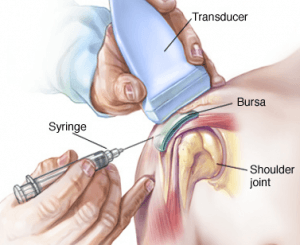
Dr. Domb and his team are highly trained in ultrasound-guided injections. These procedures are performed in the office at the time of your visit. The ultrasound machine assists Dr. Domb and his Physician Assistants to safely place injectable medications into or around the joint. Shoulder injection options include:
Cortisone:
A combination of steroid medication and local anesthetic to provide pain relief and decrease inflammation caused by an intra-articular injury or arthritis. A patient can receive a cortisone injection every 3-4 months.
- Intra-articular injections
- Subacromial injections
- Acromio clavicular joint injections
- Biceps tendon injections
Platelet Rich Plasma (PRP)
Platelet Rich Plasma (PRP) is an injection of your own platelets and growth factors, similar to orthobiologics treatment, extracted from your blood. The blood draw and injection are done at the same office visit, an outpatient clinical procedure.
PRP has shown great promise in stimulating repair of body tissues including tendons, ligaments and cartilage. PRP mimics the body's innate response to an injury by stimulating platelet activation. The activation of platelets plays a dynamic role in soft tissue healing. Research shows that PRP is superior to other injections in the treatment of osteoarthritis and healing of chronic tendinitis.
In osteoarthritis, PRP can enhance the body's normal healing response in an acute manner for a chronic condition. This can result in the development of new collagen, a benefit that other injections are unable to offer.
In tendinitis, the growth factors in PRP can signal the body to initiate a healing response in the local injected tissues. Healing is the first step in injured tissues regenerating their strength and function. It has been used extensively in professional athletes who seek hurried return to play. Star athlete success stories include: Kobe Bryant (NBA), Tiger Woods (PGA tour), Alex Rodriquez (MLB), Hines Ward (NFL) and Rafael Nadal (ATP).
If you have an injury or condition involving a tendon, ligament, joint or osteoarthritis, PRP may be a nonsurgical option for you.
Indications (including but not limited to):
- Osteoarthritis (Intraarticular)
- AC Joint
- Glenohumoral joint
- Tendonitis Tendinosis (Shoulder &Elbow)
orthobiologics Therapy
orthobiologics therapy is a form of regenerative medicine that utilizes the body’s natural healing mechanism to treat various conditions.
orthobiologics are being used in regenerative medicine to renew and repair diseased or damaged tissues and have shown promising results in treatments of various orthopedic, cardiovascular, neuromuscular and autoimmune conditions.
orthobiologics are present in all of us acting like a repair system for the body. However, with increased age sometimes the optimum amount of orthobiologics are not delivered to the injured area. The goal of orthobiologics therapy is to amplify the natural repair system of the patient’s body.
Types of orthobiologics
There are two major types of orthobiologics embryonic orthobiologics and adult orthobiologics. Embryonic orthobiologics (ESCs) are orthobiologics derived from human embryos. They are pluripotent, which means they have the ability to develop into almost any of the various cell types of the body.
As the embryo develops and forms a baby, orthobiologics are distributed throughout the body where they reside in specific pockets of each tissue, such as the bone marrow and blood. As we age, these cells function to renew old and worn out tissue cells. These are called adult orthobiologics or somatic orthobiologics. Like embryonic orthobiologics, adult orthobiologics can also replicate into more than one cell type, but their replication is restricted to a limited number of cell types.
Use of orthobiologics in Orthopedics
The unique self-regeneration and differentiating ability of embryonic orthobiologics can be used in regenerative medicine. These orthobiologics can be derived from eggs collected during IVF procedures with informed consent from the patient. However, many questions have been raised on the ethics of destroying a potential human life for the treatment of another.
Adult orthobiologics are most commonly obtained from the bone marrow, specifically the mesenchymal orthobiologics, which have the ability to replicate into cells that form the musculoskeletal system such as tendons, ligaments, and articular cartilage. They can be obtained from the iliac crest of the pelvic bone by inserting a needle and extracting the orthobiologics from the bone marrow.
Currently, orthobiologics therapy is used to treat various degenerative conditions of the shoulder, knees, hips, and spine. They are also being used in the treatment of various soft tissue (muscle, ligaments and tendons) as well as bone-related injuries.
Who is a Good Candidate for a orthobiologics Procedure?
You may be a good candidate for orthobiologics therapy if you have been suffering from joint pain and want to improve your quality of life while avoiding complications related to invasive surgical procedures.
Preparing for the Procedure
- It is important that you stop taking any non-steroidal anti-inflammatory drugs (NSAIDs) at least two weeks before your procedure.
- Preparing for a orthobiologics procedure is relatively easy and your doctor will give you specific instructions depending on your condition.
orthobiologics procedure
The procedure begins with your doctor extracting orthobiologics from your own bone marrow. Bone marrow is usually aspirated from your hip region. Your doctor will first clean and numb your hip area. A needle is then introduced into an area of your pelvic bone known as the iliac crest. Bone marrow is then aspirated using a special syringe and the sample obtained is sent to the laboratory. In the laboratory, the aspirate is spun in a machine for 10 to 15 minutes and a concentrated orthobiologics sample is separated.
Your doctor then cleans and numbs your affected area to be treated and then, under the guidance of special x-rays, injects the orthobiologics into the diseased region. The whole procedure usually takes less than one hour and you may return home on the same day of the procedure.
Post-Operative Care
- You will most likely be able to return to work the next day following your procedure.
- You will need to take it easy and avoid any load bearing activities for at least two weeks following your procedure.
- You will need to refrain from taking non-steroidal, anti-inflammatory medications (NSAIDS) for a while as this can affect the healing process of your body.
Advantages & Disadvantages
- orthobiologics therapy is a relatively simple procedure that avoids the complications associated with invasive surgical procedures.
- As orthobiologics therapy uses the cells derived from your own body it reduces the chances of an immune rejection.
Disadvantages
- The disadvantage of adult orthobiologics therapy is lack of data about its long-term effects as it is a newer evolving therapy.
Risks and complications
orthobiologics therapy is generally considered a safe procedure with minimal complications, however, as with any medical procedure, complications can occur.
Some risks factors related to orthobiologics therapy include infection as the orthobiologics may become contaminated with bacteria, viruses or other pathogens that may cause disease during the preparation process.
The procedure to either remove or inject the cells also has the risk of introducing an infection to the damaged tissue into which they are injected. Rarely, an immune reaction may occur from injected orthobiologics.
Arthroscopic Repair of the Shoulder
Arthroscopic shoulder surgery incorporates the use of very small poke-hole incisions (portals) around the joint, and the use of a specialized camera (arthroscope) among other specified arthroscopic surgical instruments. The goal of an arthroscopic surgery is to repair and restore the joint to full strength, while maintaining full range of motion. Due to the minimally invasive nature of the arthroscopic technique, damage to surrounding muscles, ligaments, tendons, nerves and blood vessels is significantly reduced, thus, less pain and rehabilitation after surgery. There is also a reduced risk of infection compared to an open technique.
Recovery
Shoulder arthroscopy is generally an outpatient procedure. The average postoperative course involves 2-6 weeks in a shoulder sling to protect the work done on the shoulder. A sling may be required for 6 weeks if the shoulder's condition requires a more extensive surgery. Most patients begin physical therapy 2-6 weeks after surgery. Patient's return to work is extremely variable after their procedure, depending on their surgery and their work type. Athletes can expect to return to sports between 6 months and 1 year after surgery. High-level athletes participate in an intense physical therapy course after surgery, gradually increasing their workout intensity.
Rotator Cuff Repair
In arthroscopic rotator cuff repair, the torn tendon is sewn back to the bone of the humerus. Dr. Domb continues to research the most advantageous methods of repair, and his evolving innovations have been published in the Journal of Bone and Joint Surgery, the most prestigious journals in Orthopedic Surgery.
Article
SLAP Repair
In arthroscopic labral repair, the torn labrum is sewn back to the edge of the glenoid socket using sutures. Dr. Domb continues to research the most advantageous methods of repair, and his innovations have been published in the journal Arthroscopy, one of the most prestigious journal in Orthopedic Surgery.
Article
Biceps Tenodesis
Biceps tenodesis is an option for those who do not have relief or improvement with a biceps tendon tear with conservative methods. Several new, procedures have been developed to repair the tendon in a minimally invasive fashion. The goal of the surgery is to re-attach the tendon to the bone. At the level of the shoulder, the biceps tendon will usually be reattached or anchored to the proximal humerus using a small screw and suture.
Publications by Dr. Domb on Shoulder Surgeries
- High Tension Double-row Footprint Repair compared with reduced Tension Single row repair Massive Rotator Cuff Tears 2008
- Biomechanical comparison of 3 Suture Anchor Configuration for repair of type II SLAP lesions 2007
- The two-step maneuver for Closed Reduction of Inferior Glenohumoral Dislocation 2006
- Clinical Follow up of throwers Undergoing Ulnar Collateral Ligament Reconstruction using the Kerlan-Jobe Orthopaedic Clinical Shoulder and Elbow score 2010
- Complications of Circular Plate Fixation for Four-Corner Arthrodesis 2007
Get Adobe ReaderYou will need the Adobe Reader to view & print these documents.

